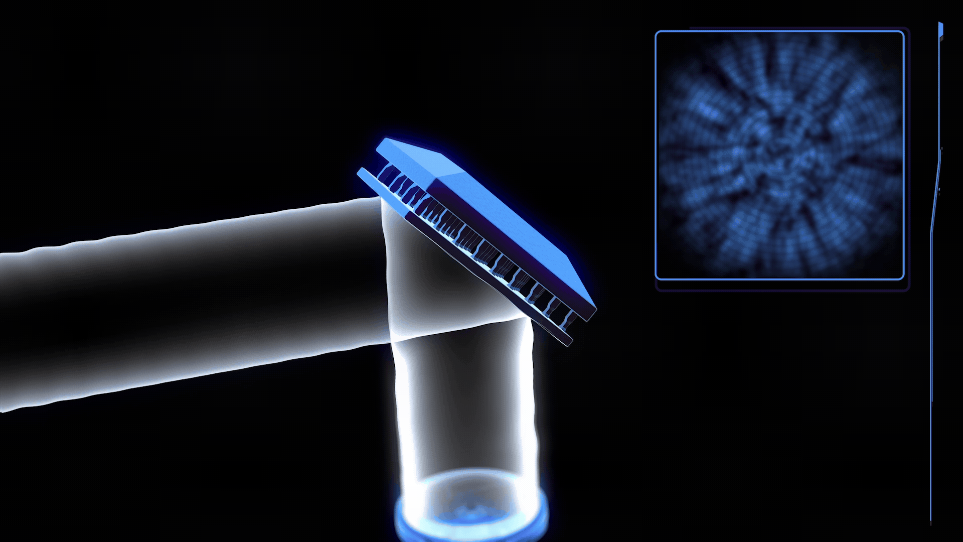

Fahy’s meticulous examination of the brain biopsies revealed an incredible level of detail. "We can see every detail [in the structure of the brain biopsies]," Fahy stated, highlighting the remarkable preservation of cellular architecture. This observation fuels his optimism that Coles’ brain might indeed stand a chance of future reanimation, a concept that has long resided in the realm of science fiction. However, not all experts share this level of optimism. John Bischof, a researcher at the University of Minnesota specializing in cryopreservation of human organs, offered a more grounded perspective: "This brain is not alive." Despite this cautionary note, Fahy’s groundbreaking research holds significant promise for neuroscientists seeking novel methods to study the brain. Furthermore, the technological advancements demonstrated in this study could pave the way for practical applications, such as the preservation of organs for transplantation, a goal that is seemingly within reach.
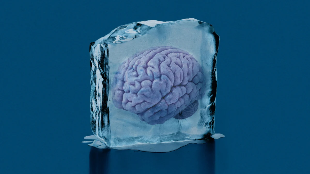
Coles’ decision to undergo cryogenic preservation was a deliberate scientific endeavor. After his death, his body was maintained at a low temperature during transport to Alcor, a renowned cryonics facility in Arizona. His head was then carefully detached, and his brain was perfused with cryoprotective chemicals designed to prevent ice crystal formation during the subsequent cooling process. This procedure, crucial for mitigating damage, involved removing the brain from the skull and gradually lowering its temperature to -146 degrees Celsius. This process, while complex, was meticulously managed, with Fahy himself involved in coordinating the extraction of the brain biopsies. Nick Llewellyn, who oversees research at Alcor, confirmed Fahy’s integral role, noting that he was "on the phone coordinating the whole thing, [including] where the biopsies were taken." These small samples were then stored in liquid nitrogen, specifically earmarked for Fahy’s research, while the rest of the brain was cooled and maintained in a temperature-controlled environment at Alcor.
The motivation behind this scientific undertaking was multifaceted. Hundreds of individuals have opted for cryogenic preservation of their brains, with or without their entire bodies, at facilities like Alcor, where the remains of 259 individuals are currently stored. However, concrete scientific understanding of the long-term effects of these procedures on brain tissue remains limited, with no definitive evidence to support the possibility of revival. Coles, driven by his scientific curiosity and his shared interest in longevity with Fahy, specifically requested his brain be studied. "He thought that if he had himself cryopreserved, we could learn from his brain whether cracking was going to happen or not," Fahy explained. This concern about cracking, a common issue when tissues are subjected to rapid cooling to ultra-low temperatures like -196 degrees Celsius (liquid nitrogen), stems from the immense tension created within the material, which can lead to shattering. Fahy noted that the slightly warmer preservation temperature of -146 degrees Celsius makes such catastrophic cracking less likely.
Fahy’s investigation into the cryopreserved brain samples, which commenced years after their extraction, focused on the potential impact of the cryoprotectant chemicals. These chemicals, while essential for preventing ice formation, are inherently toxic and can, in some instances, distort cellular structures. Previous research had indicated that flooding tissues with cryoprotectants could lead to cellular compression and structural deformation. This challenge highlights a fundamental hurdle in cryobiology: preserving delicate human tissues at extremely low temperatures without causing irreversible damage. While the vitrification of eggs and embryos, a process that cools them to -196 degrees Celsius, essentially turning them into a glass-like solid, has become relatively routine—a success partly attributed to Fahy’s pioneering work with mouse embryos in the 1980s—preserving larger, more complex organs presents a significantly greater challenge. Uniform cooling of larger objects is difficult, increasing the risk of damaging ice crystal formation and cracking, even with the use of cryoprotectants.
The results of Fahy’s rewarming and rehydration experiments were striking. He observed that Coles’ brain cells, after being rewarmed and rehydrated, exhibited a remarkable ability to "bounce back" to a degree, regaining some semblance of their original structure. Fahy vividly described this phenomenon, illustrating it with hand gestures that conveyed a transformation from a compressed state to a restored form. He assessed the tissue structure as largely intact, acknowledging that while a purist might find imperfections, the level of detail visible in the cells and their components was extraordinary. "There’s nothing we don’t see," Fahy asserted, underscoring the completeness of his observations. His findings, which have been shared on the preprint server bioRxiv and are awaiting peer review, suggest that the cryogenic approach, at least in this instance, has preserved a significant amount of cellular information. Regarding the concern of cracking, Fahy reported that the initial preservation team observed no cracks. While photographs taken by Alcor during the biopsy procedure were unfortunately lost due to a server malfunction, recent images of the brain reveal a layer of frost, making it impossible to definitively assess for cracks without risking further damage. Consequently, the team has opted to leave the frost undisturbed.
Following the rewarming process, Fahy and his colleagues employed chemical fixation techniques to stabilize the brain samples. This standard procedure, used to prevent decay in fresh tissue samples, effectively renders them non-viable in the traditional sense. However, Fahy’s research provides compelling evidence that cryopreservation of small brain tissue sections may indeed be a viable pathway for reanimation and subsequent study of their functions. Recent breakthroughs in Germany have demonstrated the revival of mouse brain slices stored at -196 degrees Celsius, with these samples exhibiting electrical activity post-cooling and rewarming. If similar results can be achieved with human brain samples, these preserved tissues could offer neuroscientists invaluable insights into the intricate workings of living brains.
Shannon Tessier, a cryobiologist at Massachusetts General Hospital, echoed this sentiment, suggesting that brain cryopreservation "can capture a little bit more of the complexities of the brain." She believes that the ability to study human brains from deceased individuals would significantly enhance the researcher’s toolkit. Matthew Powell-Palm, a cryobiologist at Texas A&M University, described Fahy’s work as providing critical information about "what happens when we try and vitrify a one-liter, dense, massive goop." He added that the research offers "a strong indication that quite large [tissues and organs] can be vitrified by perfusion [without forming too much ice]."
Beyond the realm of fundamental neuroscience, the scientists involved in this research, including Fahy, are actively pursuing the development of technologies for organ preservation for transplantation. The scarcity of transplantable organs is a significant global health challenge, exacerbated by the limited window of time an organ remains viable after removal from a donor. Cryopreservation offers a potential solution, extending the viability of organs and allowing for more efficient organ matching, preparation of recipients’ immune systems, and potentially reducing the lifelong reliance on immunosuppressant drugs, as noted by Bischof. Significant progress has already been made in this area, with successful cryopreservation and transplantation of organs in animals like rabbits and rats. Bischof expressed confidence, stating, "We’re at the cusp of human-scale organ cryopreservation."
However, the ultimate goal of brain cryopreservation, as envisioned by Coles, is reanimation—a far more ambitious objective that hinges on the ability to restore brain function. Fahy acknowledges that while the structural integrity of Coles’ brain samples has been impressive, there is currently no evidence to suggest that the cells can be brought back to life, regain electrical activity, or resume a functioning metabolism. "Restoring it to function… that’s a whole other story," he admitted. Nevertheless, Fahy sees successful cryopreservation of the brain as a crucial step toward achieving "human suspended animation," a technology that could enable long-duration space travel. He also envisions it as a form of "medical time travel," allowing individuals to avoid death by journeying to a future where cures for their ailments might exist.
The likelihood of achieving brain reanimation remains a subject of cautious optimism. Llewellyn from Alcor estimates the chances as "pretty low," emphasizing that the necessary technology is "practically unfathomable." Tessier further cautions that the methods used for cryopreserving brains currently stored in facilities have "not been validated to work for reanimation," labeling the expectation of future revival as a "quite a jump of faith and hope that’s not based on science." As Powell-Palm succinctly put it, "There are so many ways in which those neurons could be toast." Despite these formidable challenges, the study of L. Stephen Coles’ cryopreserved brain represents a significant stride, pushing the boundaries of scientific understanding and offering a glimpse into the future possibilities of cryobiology and its potential to redefine life and death.